A false positive is an alert the evidence doesn't support, and there's no single universal false positives rate for heart-rate alerts. First identify whether you're questioning the recorded episode, its confirmation on another test, or what it means medically.
You stand up from a chair, your watch flags a rise, and suddenly you're studying a graph instead of making breakfast. If you've been dealing with dizziness or palpitations, that notification can feel both reassuring and unsettling: something happened, but does the alert explain it?
You don't have to choose between trusting every notification and ignoring your watch. A better approach is to separate what the sensor recorded, what another measurement confirms, and what a clinician can conclude.
What a False Positive Means for Heart-Rate Alerts
Say your watch flags a rise after you stand up. Is the alert wrong? Not necessarily. The sensor might have misread your pulse, the software might have counted an unsuitable event, or your heart rate might have risen without establishing postural orthostatic tachycardia syndrome, or POTS.
These are different questions, and they need different checks.
Three meanings of a false positive
| Meaning | What it counts | Example | How to check |
|---|---|---|---|
| Episode-level | A flagged rise that fails the detection criterion | A standing-related rise is flagged, but the saved record doesn't support its magnitude or persistence | Compare the recorded baseline, peak and duration |
| Confirmation-level | An alert not matched by ECG or supervised testing | A watch alert has no matching ECG confirmation | Record the rhythm when possible and seek an appropriate reference test |
| Clinical-level | A real rise with a non-POTS explanation | A rise after exercise, or a misleading interpretation involving ectopic beats | Ask a clinician to review the full history |
An alert without an ECG isn't automatically false. Unconfirmed means you don't yet have enough evidence to classify it confidently.
The apparent contradictions in accuracy figures often come from mixing these meanings. In a large consumer-wearable heart study published in Circulation, only about one-third of notified participants had atrial fibrillation on subsequent one-week ECG monitoring, while the concurrent patch-monitored subgroup had a 98.2% positive predictive value and a reported 1.5% false-positive rate.
Check what the percentage divides by
Positive predictive value asks how many positive alerts were correct. A false-positive rate asks how often the system flagged a positive among observations that were negative. They use different denominators, so subtracting positive predictive value from the whole doesn't automatically give the false-positive rate.
Timing matters too. A later recording may not capture an intermittent rhythm that occurred during the notification. The condition's prevalence and which participants receive confirmation testing also affect how the results should be read.
A screening alert is like a smoke alarm: it tells you to investigate, not what caused the alarm.
Person-level results count people; episode-level results count events. Someone with repeated alerts still represents a single person in a person-level analysis.
Before judging an alert, write down your actual question: Was the measurement believable, was the event confirmed, or does it support a diagnosis?
Common Causes of False Alerts and How to Check Each
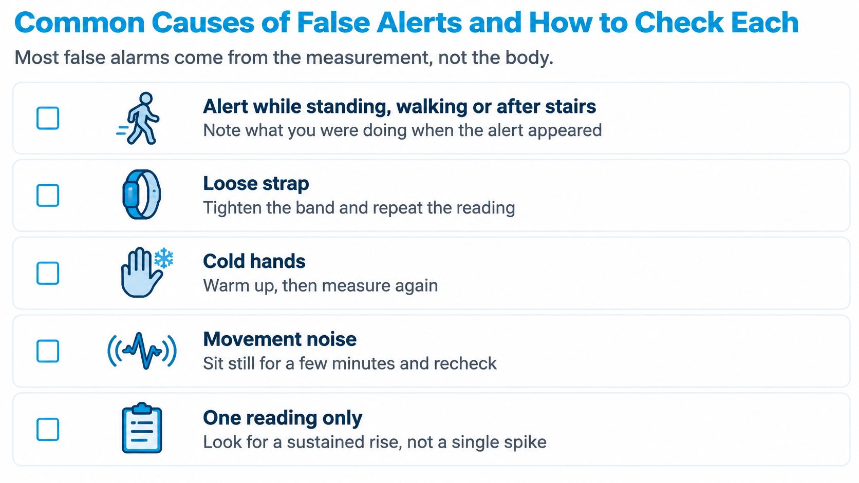
An alert appears while you're standing, walking or finishing the stairs. Before assuming that your body did something unusual, check whether your wrist was moving and whether the watch maintained good contact.
Optical wrist sensors use photoplethysmography, usually shortened to PPG, to estimate pulse from changes in reflected light. Wrist movement can shift the sensor, deform the skin and alter local blood flow. The resulting signal can resemble a pulse change even when it doesn't accurately represent your heart rate.
An independent validation discussed in this wearable-monitoring paper found that absolute heart-rate error during physical activity was, on average, 30% higher than at rest, and reported rates could be systematically higher than the true rate during activity.
That doesn't mean every moving reading is wrong. It means movement deserves its own check rather than being treated as irrelevant background.
Match the pattern to a practical check
| Cause | What it looks like | How to check |
|---|---|---|
| Arm movement or walking | Spikes that start and stop with activity | Check the activity label and timing of the rise |
| Loose or misaligned watch | Erratic, flat or jumping readings | Refit the watch snugly above the wrist bone and recheck |
| Low perfusion | Readings that lag or drop when hands are cold | Warm your hands and compare with a manual pulse count at the same moment |
| Peripheral edema or bright ambient light | A noisy signal without an obvious matching body change | Note swelling or lighting and repeat the reading with better sensor contact |
| Ectopic beats | Irregular or skipped beats you may feel | Compare the episode with an ECG strip if one is available |
The last row needs particular care. Ectopic beats aren't just a loose-strap problem, and a sensation of skipped beats doesn't establish which rhythm occurred. A pulse check can help you notice disagreement, but it can't identify the rhythm as an ECG can.
Recheck without rewriting the event
If you're comfortable and safe, stop moving, adjust the fit and take another reading. Keep the original episode rather than replacing it with the cleaner result. A normal recheck tells you about the later moment, not necessarily what happened during the alert.
Don't tighten the strap until it's uncomfortable. Aim for stable contact, and note whether the graph changed after the adjustment. Our guide to wrist-based heart-rate accuracy explains how to assess these measurement limitations.
Consumer-hardware heart-rate data can be incomplete, delayed or inaccurate, particularly during movement or poor sensor contact. An empty activity label is also not proof that you were resting.
Excluding workouts and recovery periods is useful, but it isn't a complete solution. Low perfusion, loose contact, swelling, light interference and misalignment can still affect readings outside exercise.
How Episode Length Changes Alert Reliability
A brief spike is like a sudden noise in a quiet room: you notice it, but there's little information to interpret. A sustained rise is more like a conversation you can follow. You can inspect how it begins, whether it persists and how it ends.
Duration affects both missed events and misleading detections. A watch may fail to capture a short real episode, while software that saves every isolated peak may preserve sensor noise as though it were a meaningful event.
What the duration evidence actually measures
An electrophysiology study of 52 patients with induced supraventricular tachycardia reported the duration-specific results below. These measurements concern induced SVT, not POTS, so they describe a technical limitation rather than a POTS diagnostic accuracy rate.
| Episode length | Registered increase from baseline | Agreement with ECG heart rate |
|---|---|---|
| Under 15 seconds | 18.7% | Not specified in the cited summary |
| 15 to 60 seconds | 67.6% | Not specified in the cited summary |
| 60 seconds or more | Separate detection percentage not specified | 23 of 23 episodes at 90% or higher |
The table reports different outcomes. Registering an increase means the wearable detected a change from baseline. Agreement with ECG describes how closely its heart-rate estimate matched the reference measurement. Those aren't interchangeable measures.
The absence of a separate detection percentage in the final row shouldn't be filled with an assumption. Likewise, strong agreement during longer induced episodes doesn't establish how well a watch detects standing-related symptoms in daily life.
Why a saved duration field matters
An episode record should let you distinguish a peak from a sustained pattern. If you can see only the highest value, you can't tell whether surrounding readings support it or whether the apparent rise rests on an isolated sample.
There's a trade-off. Requiring persistence helps avoid transient spikes, but an excessively long requirement can exclude short real events. Brief doesn't automatically mean false, and sustained doesn't automatically mean diagnostic.
Practical rule: Check how long the rise was supported by actual readings before treating the peak as an episode.
Our guide to episodes of tachycardia can help you distinguish the event's shape from its highest number. When reviewing your own record, look for the saved duration, gaps in sampling and the app's stated persistence rule.
If that rule isn't visible, keep the event marked as uncertain rather than inventing a cutoff yourself.
When a Real Rise Still Is Not Diagnostic
A heart-rate rise can be real and still not explain your symptoms. Accurate measurement answers whether your pulse changed; it doesn't settle why it changed or whether a particular diagnosis fits.
Even the testing setup can influence classification. An Oxford-hosted report discussing orthostatic testing summarized a 60-person study in which 74% met the heart-rate criterion during active standing, compared with 98% during head-up tilt.
That contrast doesn't tell you which result your own test would produce. It shows why a percentage without its protocol can be misleading: active standing and supported tilting aren't interchangeable situations.
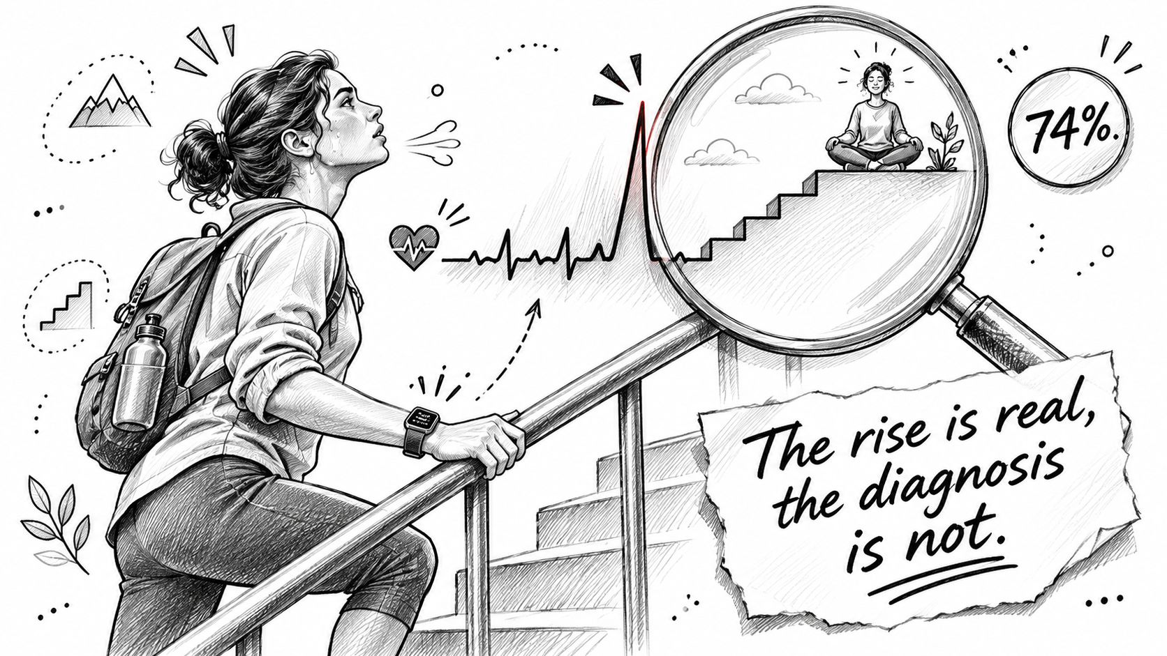
An incorrect label can conceal a real finding
Wearable rhythm findings also require interpretation. False-positive atrial-fibrillation classifications can involve other rhythms, including atrial flutter, atrial tachycardia and sinus rhythm. Premature atrial or ventricular contractions can complicate automated classification too.
The important distinction is between an incorrect disease label and nothing happening. A clinician may reject the automated label while still deciding the underlying rhythm deserves attention.
Think of a smoke alarm triggered by burnt toast. There isn't a house fire, but there is a real reason the alarm sounded. Similarly, a correctly measured rise may have an explanation other than the condition you're investigating.
Bring the circumstances into the interpretation
Useful context includes:
- Hydration and illness: Record whether you were unwell or drinking differently from usual.
- Medications: Note timing and recent changes, but don't alter prescribed treatment to test a watch reading.
- Posture and recovery: Describe whether you stood still, walked immediately or had recently exercised.
- Measurement conditions: Keep notes about sensor contact and interrupted readings separate from symptoms.
- Testing protocol: Tell the clinician whether the event came from daily activity, a home observation or supervised testing.
These notes aren't a way to diagnose yourself by elimination. They're a way to give the clinician a clearer account of what happened.
A useful record preserves uncertainty. It retains the underlying measurements, identifies patterns that might need ECG confirmation and allows comparison across days without claiming that repetition proves a cause.
Repeated observations can make a pattern easier to interpret, but they can't eliminate sensor errors or physiological confounders. A credible flagged rise tells you that heart rate moved, not why it moved.
How to Read an Episode Record Step by Step
Review an alert when you're comfortable and able to concentrate. Your aim isn't to prove it right or wrong at all costs. It's to decide what you can support, what remains uncertain and what deserves clinical review.
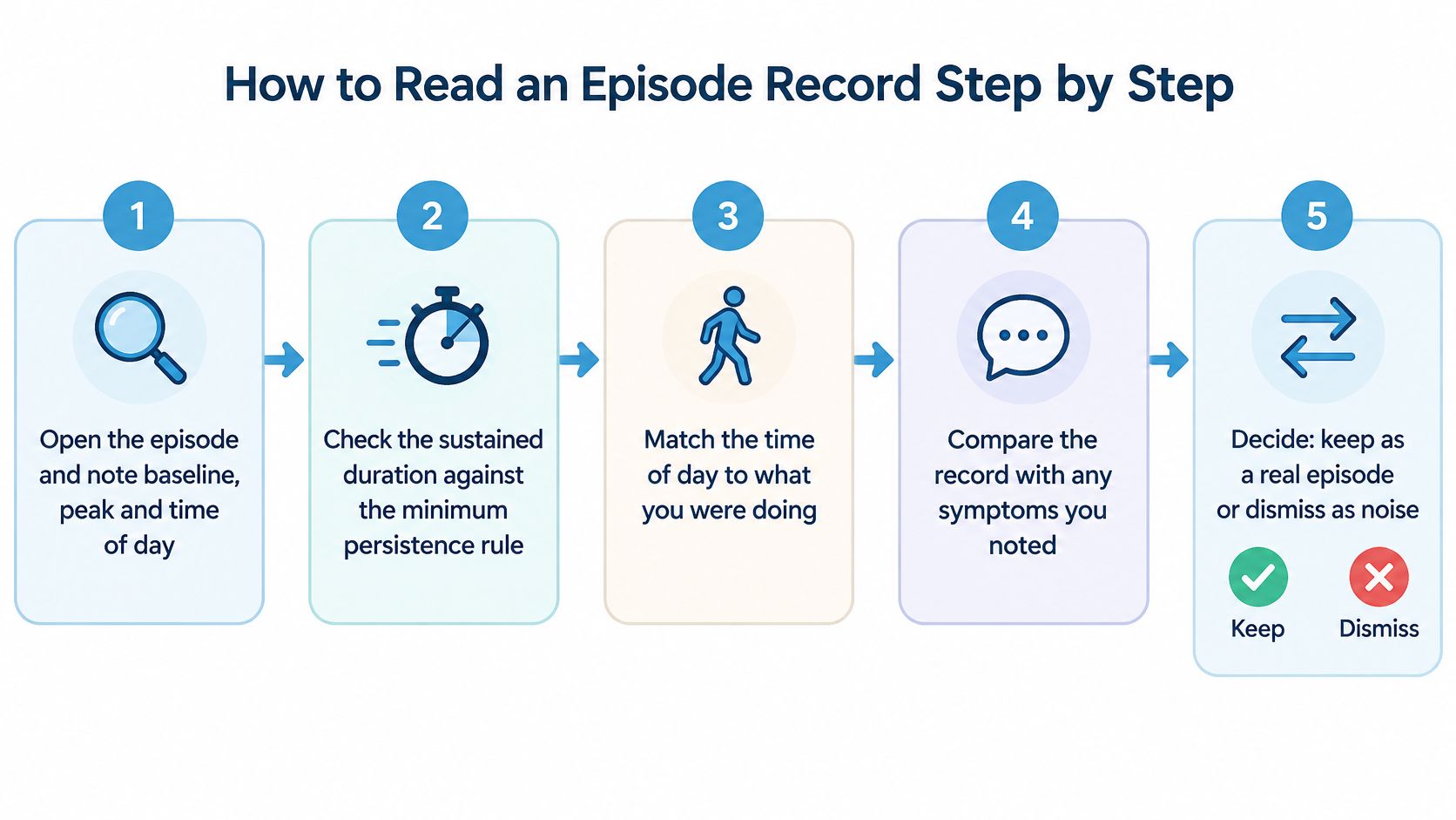
A practical review routine
Open the original episode. Note the baseline, peak and time of day so you can locate the event again. Don't save only a cropped peak, because that removes the surrounding evidence.
Check sustained duration. Compare the record with the detector's stated persistence rule and look for gaps between samples. Don't assume a line drawn between distant readings proves the rise continued throughout.
Match activity to the timestamp. Check whether walking, a workout or recovery overlapped the event. If the activity label is missing, write down what you remember rather than treating the blank as confirmed rest.
Add body and symptom context. Note posture, hydration, sleep, medication timing, illness and any irregular beats you felt. Keep observations separate from interpretations: write that you felt dizzy after standing, not that the alert proved POTS.
Inspect signal plausibility and watch fit. Look for abrupt jumps, unexpected flat stretches or changes around a strap adjustment. A tidy graph alone isn't proof of accuracy, so include what you know about contact and movement.
Compare a simultaneous second signal when available. A manual pulse can help check the rate, while an ECG strip can provide rhythm information. Don't compare a later resting measurement with an earlier symptomatic peak and call the difference an error.
Choose a provisional category. Mark the episode as likely noise, unclear or worth raising with a clinician, and add a short reason. Don't force an uncertain event into a confident yes-or-no decision.
What a clean record looks like
Say your saved event shows a steady seated baseline, followed by a sustained rise after standing. You note that you stayed still, felt lightheaded, had stable watch contact and obtained a matching pulse check while the rise was occurring.
That's a reviewable record, not a diagnosis. Its strength is that another person can understand the sequence and see which checks you performed.
Keep unclear episodes available for review. Dismissing uncertainty as noise can remove information a clinician would have wanted.
You don't need a perfect diary. A short, accurate note is more useful than a detailed account reconstructed later with false confidence.
How Context-Aware Detection Reduces Noise
Context-aware detection works best as a sequence of decisions, not a single filter that promises clean results. Each decision should have a visible purpose, and the original event should remain understandable after filtering.
A useful design separates raw crossings, candidate episodes and reviewable sustained episodes. Otherwise, a person looking at the final count can't tell whether few events occurred or whether many were removed.
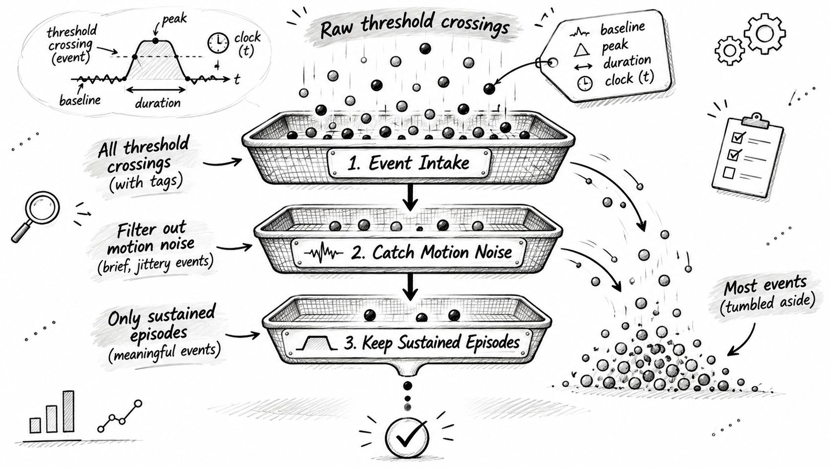
Preserve the evidence before filtering
The initial record should retain baseline, peak, duration and time of occurrence. Activity screening can then distinguish an ordinary workout response from a candidate standing-related event, rather than letting both contribute to the same unexplained-rise count.
Persistence screening serves a different purpose: it distinguishes an isolated crossing from an event supported over time. Borderline records should remain identifiable for review instead of being presented with the same confidence as well-supported episodes.
Our reference approach uses a 30 bpm rise within 5 minutes, with a 40 bpm threshold for ages 12 to 19. These thresholds are reference points, not diagnostic criteria. The intended record keeps baseline, peak, sustained duration and occurrence time, while separating workouts and recovery from episode counts.
That describes a design to validate, not proof of a particular false-positive rate. No performance percentage should be inferred from the presence of those features alone.
Make exclusions visible
A useful summary should distinguish why candidate events weren't included:
- Exercise exclusions: The rise overlapped a workout.
- Recovery exclusions: The rise occurred during post-exercise recovery.
- Duration exclusions: The record didn't meet the stated persistence requirement.
- Signal-quality concerns: Missing or implausible readings limited interpretation.
- Review-needed events: The available context didn't support confident classification.
This information supplies the missing denominator. A low final count could reflect few initial crossings, extensive activity exclusions or poor-quality recordings. Those situations shouldn't look identical to the reader.
For validation, the important question is how the detector performs at rest, during posture transitions, during ordinary movement and during exercise. A combined accuracy figure can conceal weaker performance in the setting where a particular person receives most alerts.
The reference comparison also matters. Episode-level validation should establish whether a flagged event matches an appropriate ECG or supervised orthostatic measurement, not merely whether average heart rates look similar.
Fewer notifications aren't automatically better detection. Filters should remove unsuitable events without hiding their reasons or encouraging the reader to interpret silence as proof that nothing happened. The remaining flags still deserve a human check.
Bring Flagged Episodes to Your Next Appointment
Bring selected episodes to a clinician rather than treating a notification as a verdict. Choose records that represent your symptoms or recurring pattern, including an unclear example if it captures something you're struggling to explain.
Prepare each with baseline, peak, duration, activity and symptoms, plus notes about checks you performed. If an exported report is available, include it rather than relying on disconnected screenshots.
Questions that make the appointment more useful
- Reliability: Which of these records do you consider interpretable?
- Confirmation: Which patterns need ECG confirmation?
- Next testing: What assessment would help confirm or rule out the suspected explanation?
A report is a record to discuss, not an answer by itself. Our PDF format shows episodes, trends and symptoms together and states the exact criterion used, so the reviewer can see what triggered the classification. For help organizing the conversation, use our guide to patient-provider communication.
Seek urgent care for chest pain, fainting, or symptoms that are severe or persistent. Don't wait for another notification, a report or a scheduled appointment to decide whether to seek help.
Before your visit, export or print the record and mark which episodes you've already checked using the review routine. Leave uncertain events labeled uncertain, and write your most important question at the top.
Cardiogram offers automatic episode detection, context logging and clinician-ready PDF reports to help you turn heart-rate alerts into records you can review. Visit Cardiogram to explore how episodes, symptoms and trends can support a more informed conversation with your clinician.